Products / Topography

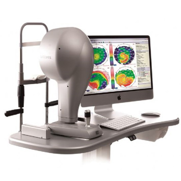
CSO Antares
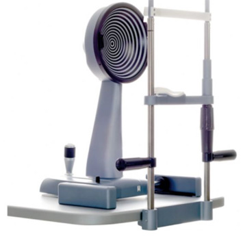
CSO Modi 02
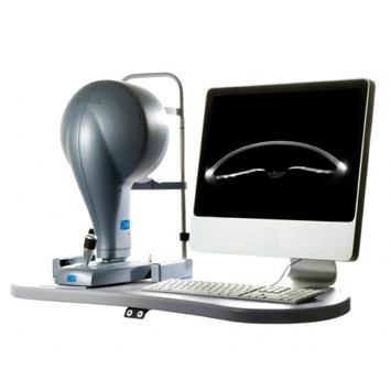
CSO Sirius
CSO ABERROMETER OSIRIS
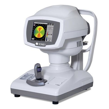
TOMEY AUTO REF- TOPOGRAPHER RT-7000
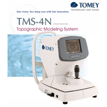
TOMEY TMS-4N Topographic Modeling System
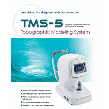
TOMEY TMS-5 Topographic Modeling System
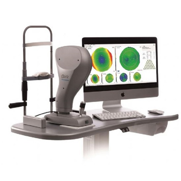
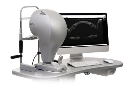
CSO CSO MS-39

  • CSO Antares

    ANTARES is the new corneal topographer that includes a new color high resolution digital camera to perform an advanced tear-film analysis. Available functions are: topography, pupillography, BUT analysis and MGD analysis. It works with USB3 interface and Phoenix software.

    Brochure

  • CSO Modi 02

    Latest branch on the CSO topographer tree, the corneal topographer. Modì02 - completely based on Firewire digital acquisition technology - is particularly suited for novices to corneal topography. With the new software Phoenix, without its advanced features, it however maintains all the basic functionality necessary for an accurate diagnosis.

    Brochure

  • CSO Sirius

    Sirius is a high precision instrument for the tomography of the anterior ocular segment and the 3D cornea analysis. By merging Scheimpflug technology, which allows the measurement of the internal ocular structures, with Placido topography, still unsurpassed for the measurement of elevation and curvatures, Sirius proves to be an essential tools for the diagnosis and the pre- and post-operative evaluation of cataract and refractive surgery.

    Brochure

  • CSO ABERROMETER OSIRIS

    OSIRIS is an aberrometer with unrivalled performance, based on a high-resolution pyramid wavefront sensor. Optical characteristics of the cornea and entire eye are analysed in a single measuring procedure. Ocular wavefront aberrations are evaluated by processing 45000 measuring points, in real time, ensuring a new level of details resolution and precision.


  • TOMEY AUTO REF- TOPOGRAPHER RT-7000

    The Auto Ref-Topographer RT-7000 is a 3-in-1 hybrid system consisting of an Auto Refractmeter, an Auto Keratometer and a Corneal Topographer. The most remarkable clinical applications of the RT-7000 are the “Tear Stability Analysis System (TSAS)” and the “Fourier Analysis map”. The TSAS interprets the changes of the mire rings’ distortion on an eye opened for 6 seconds as changes of its tear layer status and displays color code maps to illustrate the locations of those changes. On the other hand, the newly implemented “Fourier Analysis map” performs Fourier analysis for refractive powers at individual measurement points obtained by analyzing Mire images within the ø3 mm range and ø6 mm range, and visually and quantitatively displays the constant term (the spherical component), the first order term (the asymmetry component), the second order term (the regular astigmatism component), and the higher order terms (the higher order irregularity).

    Brochure

  • TOMEY TMS-4N Topographic Modeling System

    The Topographic Modeling System TMS-4 is a device that provides information about corneal configuration and refractive characteristics. Unlike conventional keratometers, this device employs color maps incorporating the most modern computer technology, to permit easier and more accurate diagnosis than conventional devices. The TMS-4 has a “Light Cone” keratoscope with a short working distance, therefore it is easier for the operator to lift the patient’s eyelid.

    Remarkable features of the TMS-4 are the auto shot system that facilitates operation and the wide variety of clinical applications developed in the long history of the TMS series.

    Brochure

  • TOMEY TMS-5 Topographic Modeling System

    The Topographic Modeling System TMS-5 is a hybrid anterior segment analyzing system consisting of a Placido ring topographer “TMS” and a 3D Scheimpflug camera. The TMS-5 is indicated for cross sectional imaging of anterior segment components of the human eye such as the anterior and posterior surface of the cornea and the anterior chamber, and also for dimension measurements of these such as curvature, length and area by computed analysis. The prominent features of the TMS-5 are the high speed Scheimpflug camera that takes only 1.0 sec. /exam, which can be very beneficial in preventing motion artifacts, and a high speed computer processing algorithm that reduces the time it takes to delineate the images.

    Brochure

  • CSO CSO MS-39

    MS-39

    AS-OCT

    Is the most advanced device for the analysis of the anterior segment of the eye. MS-39 combines Placido disk corneal topography, with high resolution OCT-based anterior segment tomography. The clarity of the cross-sectional images, with a 16 mm diameter, along with the many details of the cornea structure and layers revealed by the MS-39, will be appreciated by  anterior segment specialists. MS-39 provides information on pachymetry, elevation, curvature and dioptric power of both corneal sufaces.

    In addition to anterior segment clinical diagnostics, MS-39 can be used in corneal surgery for refractive surgery planning. An IOL calculation module is also available, based on Ray-Tracing techniques, Additional tools allow MS-39 to perform accurate pupil diameter measuremets and the advanced analysis of tear film.

    Brochure
 Back to Top